
This laboratory manual serves as a comprehensive guide for exploring human anatomy and physiology through hands-on experiments and detailed studies‚ fostering practical understanding of bodily systems and functions.
1.1. Overview of the Laboratory Manual
The laboratory manual provides a detailed and structured approach to studying human anatomy and physiology through interactive and practical exercises. It is designed to complement theoretical knowledge with hands-on experiences‚ enabling students to explore complex biological systems in a controlled environment. The manual is divided into sections‚ each focusing on specific body systems‚ such as the skeletal‚ muscular‚ digestive‚ respiratory‚ circulatory‚ nervous‚ and urinary systems. It includes visual aids‚ step-by-step procedures‚ and activities to enhance understanding and retention of key concepts.
1.2. Importance of Laboratory Studies in Anatomy & Physiology
Laboratory studies are crucial for understanding the intricacies of human anatomy and physiology. They provide hands-on experiences that complement theoretical knowledge‚ allowing students to visualize and explore biological structures and functions. Practical experiments enhance comprehension of complex systems‚ fostering critical thinking and scientific inquiry. Labs also develop essential skills in observation‚ measurement‚ and analysis‚ preparing students for real-world applications in healthcare and research. By engaging directly with specimens and models‚ learners gain a deeper appreciation of the body’s systems and their inter relationships.
1.3. Safety Protocols in the Lab
Adhering to safety protocols is essential in anatomy and physiology labs to ensure a secure environment for all participants. Proper use of personal protective equipment (PPE)‚ such as gloves and lab coats‚ is mandatory. Students must follow strict hygiene practices‚ including handwashing and surface disinfection. Handling of biological specimens and chemicals requires careful attention to avoid accidents. Emergency procedures‚ like fire extinguisher locations and spill cleanup‚ should be understood by all. Observing these guidelines prevents potential hazards and ensures a safe‚ productive learning experience for everyone involved.

Setting Up the Laboratory Environment
Setting up the lab involves organizing essential equipment‚ preparing specimens‚ and maintaining a clean environment. Proper arrangement ensures efficiency and safety during experiments and studies.
2.1. Essential Equipment for Anatomy & Physiology Labs
The anatomy and physiology lab requires specific equipment to ensure effective learning and experimentation. Essential tools include microscopes for cellular studies‚ dissecting kits with scalpels and forceps‚ and skeletons or bone models for understanding structural anatomy. Additional items like heart and lung models‚ nerve charts‚ and specimen jars are crucial for hands-on exploration. Lab coats‚ gloves‚ and goggles are vital for safety. These resources collectively provide a comprehensive environment for analyzing human systems and conducting experiments‚ fostering practical understanding and skill development in anatomy and physiology.
2.2. Preparation of Specimens and Models
Preparing specimens and models is crucial for effective anatomy and physiology lab work. Specimens are typically preserved through methods like formaldehyde fixation or freezing to maintain structural integrity. Models‚ such as 3D representations of organs or systems‚ are labeled to highlight key features. Proper storage and handling ensure longevity and safety. These preparations enable students to study complex anatomical structures and physiological processes in detail‚ fostering a deeper understanding of human body systems through hands-on examination and interactive learning experiences.
2.3. Maintaining Sterility and Hygiene in the Lab
Maintaining sterility and hygiene in the anatomy and physiology lab is essential to prevent contamination and ensure safe handling of specimens and equipment. Students should wash hands thoroughly before starting lab work and wear appropriate PPE‚ such as gloves and lab coats. Work surfaces must be disinfected‚ and instruments sterilized using autoclaves or chemical disinfectants. Proper waste disposal protocols should be followed to avoid biohazard exposure. Adhering to these practices minimizes risks and promotes a clean‚ efficient learning environment for all participants.

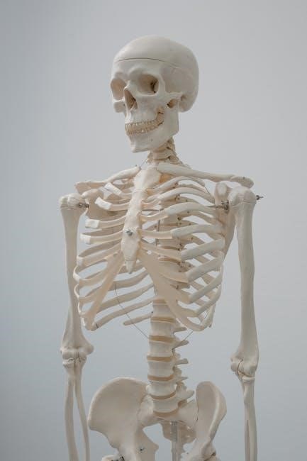
Exploring the Skeletal System
The skeletal system consists of bones‚ joints‚ and ligaments‚ providing structural support‚ protection‚ and facilitating movement while producing blood cells.
3.1. Identifying Bones and Their Functions
Bones are rigid‚ calcified structures that form the framework of the body. They vary in shape and size‚ with functions including support‚ protection‚ and movement. Long bones‚ like the femur‚ support body weight and facilitate movement‚ while flat bones‚ such as the skull‚ protect vital organs. Irregular bones‚ like vertebrae‚ provide structural support and protect the spinal cord. Each bone’s unique structure adapts to its specific role‚ enabling the skeletal system to perform essential functions for overall bodily integrity and mobility.
3.2. Axial vs. Appendicular Skeleton: Key Differences
The axial skeleton includes the skull‚ vertebral column‚ ribcage‚ and sternum‚ primarily functioning to protect vital organs like the brain and organs in the thoracic cavity. In contrast‚ the appendicular skeleton comprises the upper and lower limbs‚ shoulders‚ and pelvis‚ enabling movement and locomotion. Axial bones are generally shorter and more robust‚ while appendicular bones are longer and more specialized for mobility. Together‚ they form a cohesive system‚ with the axial skeleton providing stability and the appendicular skeleton allowing for dynamic movement and interaction with the environment.
3.3. Practical Exercises for Skeletal System Analysis
Practical exercises for skeletal system analysis involve hands-on activities to identify and study bones and their structures. Students often work with skeletal models and diagrams to label and describe bones‚ such as the cranium‚ vertebrae‚ and limb bones. Comparative studies between axial and appendicular bones are common. Exercises may include measuring bone density‚ observing joints‚ and palpating anatomical landmarks on living subjects. These activities enhance understanding of skeletal anatomy and its functional role in movement and support‚ preparing students for advanced studies in anatomy and physiology.

Muscles and Movement
This section explores the structure and function of muscles‚ including skeletal‚ smooth‚ and cardiac types‚ and their roles in movement and bodily processes through hands-on lab experiments.
4.1. Types of Muscles and Their Roles
There are three primary types of muscles: skeletal‚ smooth‚ and cardiac. Skeletal muscles‚ attached to bones‚ enable voluntary movement and support posture. Smooth muscles‚ found in internal organs like the digestive tract‚ function involuntarily‚ aiding processes like digestion. Cardiac muscle‚ exclusive to the heart‚ ensures continuous‚ rhythmic contractions for blood circulation. Each muscle type has distinct structural and functional adaptations‚ allowing them to perform specialized roles essential for maintaining bodily functions. Lab activities‚ such as dissections and microscope observations‚ provide hands-on insights into their unique characteristics and importance in human physiology.
4.2. Muscle Anatomy and Physiology
Muscles are composed of muscle fibers‚ tendons‚ and ligaments‚ working together to enable movement and stability. Skeletal muscles contract voluntarily‚ while smooth and cardiac muscles function involuntarily. Muscle contraction occurs due to the sliding filament mechanism‚ where actin and myosin filaments interact. Motor units‚ consisting of a motor neuron and its associated fibers‚ regulate contraction strength. Energy for contraction is derived from ATP‚ with glycogen serving as a reserve. Lab studies involve histological examinations of muscle tissue and experiments to observe contraction dynamics‚ providing insights into muscle physiology and its role in movement and bodily functions.
4.3. Laboratory Experiments on Muscle Function
Laboratory experiments on muscle function involve hands-on activities to observe and measure muscle contraction‚ strength‚ and fatigue. Students often perform grip strength tests‚ muscle stimulation exercises‚ and electromyography (EMG) simulations to analyze muscle activity. These experiments demonstrate how muscles respond to stimuli‚ such as electrical currents or voluntary contractions. By using tools like force transducers and data analysis software‚ learners gain practical insights into muscle physiology. Such labs enhance understanding of muscle function‚ enabling students to correlate theoretical concepts with real-world observations and applications in health and exercise sciences.
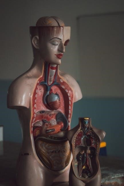
The Digestive System
The digestive system is a complex network of organs and processes that break down food‚ absorb nutrients‚ and expel waste‚ crucial for maintaining overall health and function.
5.1. Anatomy of the Upper Gastrointestinal Tract
The upper gastrointestinal (GI) tract includes the mouth‚ esophagus‚ stomach‚ and duodenum. The mouth initiates digestion with teeth and enzymes in saliva. The esophagus transports food via peristalsis to the stomach‚ where gastric juices break down nutrients. The stomach lining secretes mucus to protect itself from acid. The duodenum‚ the first part of the small intestine‚ continues digestion with bile and pancreatic enzymes. This section focuses on the structural and functional anatomy enabling food breakdown and nutrient absorption‚ forming the foundation of digestive physiology.
5.2. Physiology of the Lower Gastrointestinal Tract
The lower gastrointestinal (GI) tract includes the small intestine‚ colon‚ rectum‚ and anus. The small intestine absorbs most nutrients‚ with specialized cells and villi enhancing absorption efficiency. Enzymes and bile break down carbs‚ proteins‚ and fats into glucose‚ amino acids‚ and fatty acids. The colon absorbs water‚ solidifying waste‚ while the rectum stores feces until elimination. This process ensures proper digestion‚ nutrient uptake‚ and waste management‚ maintaining bodily function and health.
5.3. Lab Activities for Digestive System Study
Lab activities involve hands-on exploration of the digestive system‚ such as simulating digestion processes using enzymes and acids. Students analyze the role of bile salts in fat absorption and observe histological slides of intestinal tissues. Practical exercises include identifying digestive organs in models and conducting pH tests to understand stomach acidity. These activities enhance understanding of nutrient absorption‚ motility‚ and waste elimination‚ providing a dynamic learning experience for students studying anatomy and physiology.
Respiratory System
This section explores the respiratory system through detailed laboratory exercises‚ focusing on its structure‚ function‚ and role in gas exchange‚ preparing students for advanced physiological studies.
6.1. Structure and Function of the Respiratory Organs
The respiratory system consists of organs such as the nose‚ trachea‚ bronchi‚ and lungs‚ which work together to facilitate breathing and gas exchange. The nasal cavity warms and humidifies air‚ while the trachea and bronchi act as passageways for air to reach the alveoli in the lungs‚ where oxygen diffuses into the bloodstream and carbon dioxide is expelled. Laboratory exercises‚ such as dissecting lung specimens and observing alveolar structure‚ provide hands-on insights into the intricate mechanisms of respiration and the vital role of each organ in maintaining oxygenation of the body.
6.2. Breathing Mechanisms and Gas Exchange
Breathing involves the inhalation and exhalation of air‚ regulated by the diaphragm and intercostal muscles. Inhalation expands the lungs‚ drawing air into the alveoli‚ where gas exchange occurs. Oxygen diffuses into the bloodstream‚ binding to hemoglobin‚ while carbon dioxide is expelled. Laboratory experiments‚ such as measuring lung capacity and observing alveolar tissue under a microscope‚ help visualize these processes. Understanding the mechanics of breathing and gas exchange is crucial for appreciating respiratory health and function.
6.3. Hands-On Experiments for Respiratory System
Hands-on experiments in the respiratory system lab include simulating gas exchange using artificial lungs‚ measuring tidal volume with spirometry‚ and observing the diaphragm’s role in breathing. Students also explore how pH levels affect breathing rate and simulate conditions like asthma to understand respiratory challenges. These experiments provide a practical understanding of how the respiratory system functions‚ emphasizing the relationship between structure and function while reinforcing key physiological concepts through interactive learning.

Circulatory System
The circulatory system‚ comprising the heart‚ blood vessels‚ and blood‚ is essential for transporting oxygen‚ nutrients‚ and waste products throughout the body‚ maintaining homeostasis and overall health.
7.1. Heart Anatomy and Blood Vessel Types
The heart consists of four chambers: the right and left atria‚ and the right and left ventricles‚ separated by a septum. Valves ensure blood flows in one direction. Blood vessels include arteries‚ which carry oxygenated blood away from the heart (except the pulmonary artery)‚ veins‚ which return deoxygenated blood (except the pulmonary veins)‚ and capillaries‚ where nutrient and waste exchange occurs. In lab‚ students study heart dissections and histological slides of blood vessels to understand their structure and function‚ enhancing their grasp of circulatory physiology.
7.2. Blood Circulation and Pressure Measurement
Blood circulation is regulated by the heart‚ blood vessels‚ and blood pressure. In the lab‚ students measure blood pressure using sphygmomanometers and stethoscopes to understand systolic and diastolic pressures. Exercises involve analyzing blood flow through simulations and observing how factors like exercise or stress impact circulation. Practical activities include assessing pulse rates and venous return to grasp the dynamics of blood movement. This hands-on approach helps students correlate theoretical concepts with real-world physiological processes‚ enhancing their understanding of cardiovascular health and its clinical implications.
7.3. Practical Labs for Circulatory System Study
Practical labs in the circulatory system involve hands-on activities to study blood flow and vessel structure. Students perform blood pressure measurements‚ analyze blood vessel specimens‚ and simulate blood circulation using models. Additional exercises include observing blood smear slides under microscopes and conducting venipuncture simulations. These labs provide a deeper understanding of how blood moves through arteries‚ veins‚ and capillaries‚ and how factors like heart rate and vessel diameter influence circulation. Such experiments bridge theoretical knowledge with real-world applications‚ enhancing comprehension of cardiovascular physiology and its clinical relevance.
Nervous System
This section examines the structure and function of the nervous system‚ including the central and peripheral divisions‚ neural signaling mechanisms‚ reflex actions‚ and practical lab investigations;
8.1. Structure of the Central and Peripheral Nervous System
The central nervous system (CNS) includes the brain and spinal cord‚ serving as the control center for bodily functions. The peripheral nervous system (PNS) comprises nerves connecting the CNS to limbs and organs. The CNS processes information‚ while the PNS transmits sensory and motor signals. Key structures like neurons‚ glial cells‚ and myelin sheaths facilitate neural communication. Laboratory exercises often involve studying nerve tissue slides‚ dissecting specimens‚ and tracing neural pathways to understand the intricate organization of these systems and their roles in maintaining homeostasis and enabling voluntary movements.
8.2. Neural Transmission and Reflexes
Neural transmission involves the propagation of electrical and chemical signals across neurons‚ enabling communication within the nervous system. Action potentials trigger the release of neurotransmitters‚ which bind to receptors on adjacent neurons or effector cells. Reflexes are automatic responses to stimuli‚ such as withdrawing a hand from heat. Simple reflexes involve direct pathways‚ while complex ones require higher-level processing. Laboratory exercises often include studying nerve impulse conduction‚ simulating synaptic transmission‚ and observing reflex actions to demonstrate these fundamental physiological processes and their role in rapid‚ coordinated responses.
8.3. Laboratory Exercises for Nervous System Exploration
Laboratory exercises for nervous system exploration include hands-on activities to study neural structures and functions. Students often perform dissections of sheep or rat brains to identify key anatomical features. Reflex testing‚ such as knee-jerk responses‚ demonstrates automatic nervous system reactions. Additionally‚ activities involve simulating nerve impulse transmission using specialized software or models. These exercises provide practical insights into neural pathways‚ synapses‚ and the integration of sensory and motor functions‚ enhancing understanding of how the nervous system controls body responses and maintains homeostasis.
Urinary System
The urinary system‚ comprising kidneys‚ ureters‚ bladder‚ and urethra‚ plays a vital role in filtering waste‚ regulating electrolytes‚ and maintaining acid-base balance‚ essential for overall health and homeostasis.
9.1. Kidney Function and Urine Formation
The kidneys play a crucial role in filtering blood to remove waste products‚ excess water‚ and electrolytes‚ forming urine. This process involves glomerular filtration‚ where blood passes through the glomerulus and Bowman’s capsule‚ followed by tubular reabsorption and secretion. The renal corpuscle initiates filtration‚ while the renal tubules adjust the composition of the filtrate‚ regulating electrolyte balance and water levels. Hormones like aldosterone and ADH fine-tune these processes‚ ensuring proper fluid balance and electrolyte homeostasis in the body.
9.2. Anatomy of the Urinary Tract
The urinary tract consists of the kidneys‚ ureters‚ bladder‚ and urethra. The kidneys‚ located in the retroperitoneal space‚ produce urine that flows through the ureters to the bladder for storage. The bladder‚ a hollow muscular organ‚ expands to hold urine and contracts during urination. The urethra‚ a tube leading from the bladder to the exterior‚ facilitates urine expulsion. In males‚ the urethra is longer and passes through the prostate gland and penis‚ while in females‚ it is shorter and exits anterior to the vagina. This system works together to store and release urine efficiently.
9;3. Lab Activities for Urinary System Study
Lab activities for studying the urinary system include dissection of kidney models‚ microscopic examination of kidney tissue‚ and analysis of urine samples to understand filtration processes. Students may also conduct experiments to measure specific gravity and pH levels in urine‚ simulating kidney function. Additionally‚ hands-on exercises using anatomical diagrams and 3D models help visualize the urinary tract’s structure. These practical exercises enhance understanding of how the kidneys‚ ureters‚ bladder‚ and urethra work together to maintain homeostasis‚ providing a comprehensive learning experience.
The laboratory manual concludes by summarizing key concepts and encouraging students to apply their skills in real-world scenarios‚ fostering continuous learning and professional growth in anatomy and physiology.
10.1. Review of Key Concepts in Anatomy & Physiology
This section provides a comprehensive review of major concepts covered in the laboratory manual‚ emphasizing the relationship between anatomical structures and physiological functions. It highlights key themes such as the skeletal‚ muscular‚ and nervous systems‚ as well as the digestive‚ circulatory‚ and urinary systems. Practical applications of laboratory techniques are also revisited‚ ensuring a solid foundation for understanding human anatomy and physiology. The review underscores the importance of integrating theoretical knowledge with hands-on experimentation to grasp complex biological processes effectively.
10.2. Applying Lab Skills in Real-World Scenarios
Lab skills acquired through anatomy and physiology studies are invaluable in real-world applications‚ such as medical diagnostics‚ research‚ and healthcare. Techniques like dissection‚ microscopy‚ and physiological measurements are essential for professionals in these fields. Understanding bodily systems enables accurate diagnoses and effective treatments. Practical experience with lab equipment and procedures prepares individuals for careers in medicine‚ physical therapy‚ and biomedical engineering. These skills also foster critical thinking and problem-solving‚ which are crucial in addressing complex health challenges and advancing medical knowledge.
10.3. Continuous Learning in Anatomy & Physiology
Continuous learning in anatomy and physiology is essential due to the ever-evolving nature of medical research and technological advancements. Engaging in workshops‚ online courses‚ and subscribing to scientific journals helps stay updated. Collaborative learning through discussions and case studies enhances understanding. Applying knowledge in real-world scenarios‚ such as clinical practice or research‚ reinforces concepts and fosters innovation. Lifelong learning ensures professionals remain competent and adaptable in their fields‚ contributing to improved patient care and scientific progress.